

Private Hip and Pelvis MRI in Leeds: Joint, Groin & Pelvic Pain
Hip and groin pain is notoriously hard to pin down. The joint itself, the labrum, the tendons around it, the pelvic bones and even the lower spine can all produce similar symptoms. MRI shows the cartilage, labrum, bone, tendons and muscles in one examination.
This guide explains the difference between a hip MRI and a pelvis MRI, what each can show, which one is likely to be right for your symptoms, and how to arrange a private scan in Leeds.
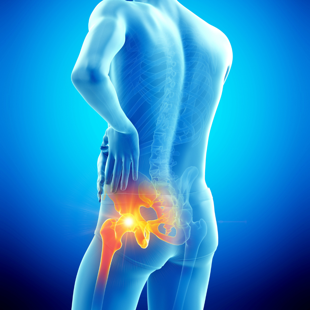
Two scans. Different questions.
The hip is a deep ball-and-socket joint surrounded by some of the strongest muscles and tendons in the body. It sits within the pelvis, a ring of bone that also contains the sacroiliac joints, the pubic symphysis and the attachments of the abdominal, groin and hamstring muscles.
A hip MRI zooms in on one joint with high-detail images. A pelvis MRI takes a wider view of both hips and the bony ring, which is better when the problem could be anywhere in the region or on both sides.
Put simply: if you know the pain is in one hip joint, a dedicated hip MRI gives the most detail. If the pain is in the groin, buttock, pelvis or both sides, or the cause is unclear, a pelvis MRI — sometimes with a hip MRI added — is often the better starting point.
What do hip and pelvis MRI scans cover?
Each examination is tailored to the clinical question, but the three most common areas of interest are set out below.
Hip MRI
Detailed imaging of a single hip: the labrum, articular cartilage, femoral head and neck, the bone marrow, the joint capsule and the tendons and bursae around the joint. Used for labral tears, impingement, early arthritis, stress fractures and avascular necrosis.
Pelvis MRI
Both hips together with the pelvic bones, pubic symphysis, muscles and tendon attachments. Used for groin strain and athletic pubalgia, hamstring and adductor injuries, pelvic stress fractures, bursitis and comparing one side with the other.
Sacroiliac joints
The two joints where the base of the spine meets the pelvis. MRI is the main test for inflammation here (sacroiliitis), which can be an early sign of inflammatory back conditions, and for mechanical SI joint problems.
When is a hip or pelvis MRI usually recommended?
An X-ray is often the first test for the hip because it shows arthritis and fractures well. MRI is requested when the X-ray is normal but symptoms persist, when soft-tissue injury is suspected, or when the result would change treatment.
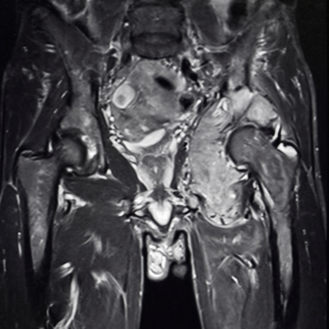
MRI for labral tears and hip impingement
The labrum is a ring of cartilage around the rim of the hip socket. Tears are a common cause of groin pain in active adults, particularly where the shape of the ball or socket causes the two to pinch during movement — known as femoroacetabular impingement, or FAI.
MRI can show the labrum, the cartilage lining the joint, bone bruising and the cysts that develop with long-standing impingement. It helps a specialist decide between physiotherapy, injection or arthroscopic surgery.
A standard hip MRI needs no injection. Where a surgeon specifically requests an MR arthrogram, with contrast injected into the joint for extra labral detail, a referral is required and this is arranged separately.
Stress fractures and avascular necrosis
Two of the most important hip diagnoses often do not show on a plain X-ray until late. MRI picks both up early.
Stress fractures of the femoral neck or pelvic bones develop with repetitive load — long-distance running, a sudden increase in training, or reduced bone density. Caught early, they heal with rest; missed, a femoral neck stress fracture can progress to a full fracture.
Avascular necrosis (AVN) is loss of blood supply to the ball of the hip. It is linked to high-dose steroids, excess alcohol, some medical conditions and previous injury. MRI is the most sensitive test and early diagnosis widens the treatment options.
Hip MRI or pelvis MRI?
Symptoms alone do not always identify the right scan, and MRI findings need to be read alongside a clinical examination. These patterns are a useful starting point, and our clinical team can confirm the choice when you book.
Hip or back? Pain in the buttock and down the thigh is often assumed to be the hip but can come from the lumbar spine, and a hip problem can be felt as knee pain. If your clinician is unsure, they may request a lumbar spine MRI alongside the hip, or a pelvis scan to cover more ground. Provide your referral or describe your symptoms when booking and our team will help you choose.
What happens during a hip or pelvis MRI?
Before your scan you will complete MRI safety screening and the radiographer will confirm the area being examined and the reason for the scan.
You lie on your back on the scanner table with your legs straight and feet gently supported so the hips stay still. A flat receiver coil is placed over the pelvis, and the table moves into the scanner feet first. For most hip and pelvis scans your head stays near the opening of the magnet, which many patients find far more comfortable than expected.
A hip MRI takes around 20 to 30 minutes; a pelvis MRI around 30 minutes, and a little longer if both are requested together. Hearing protection is provided, and staying still is the single most important thing you can do for a clear result.
You can eat and drink normally beforehand and take your usual medication. There is no injection for a standard hip or pelvis MRI.
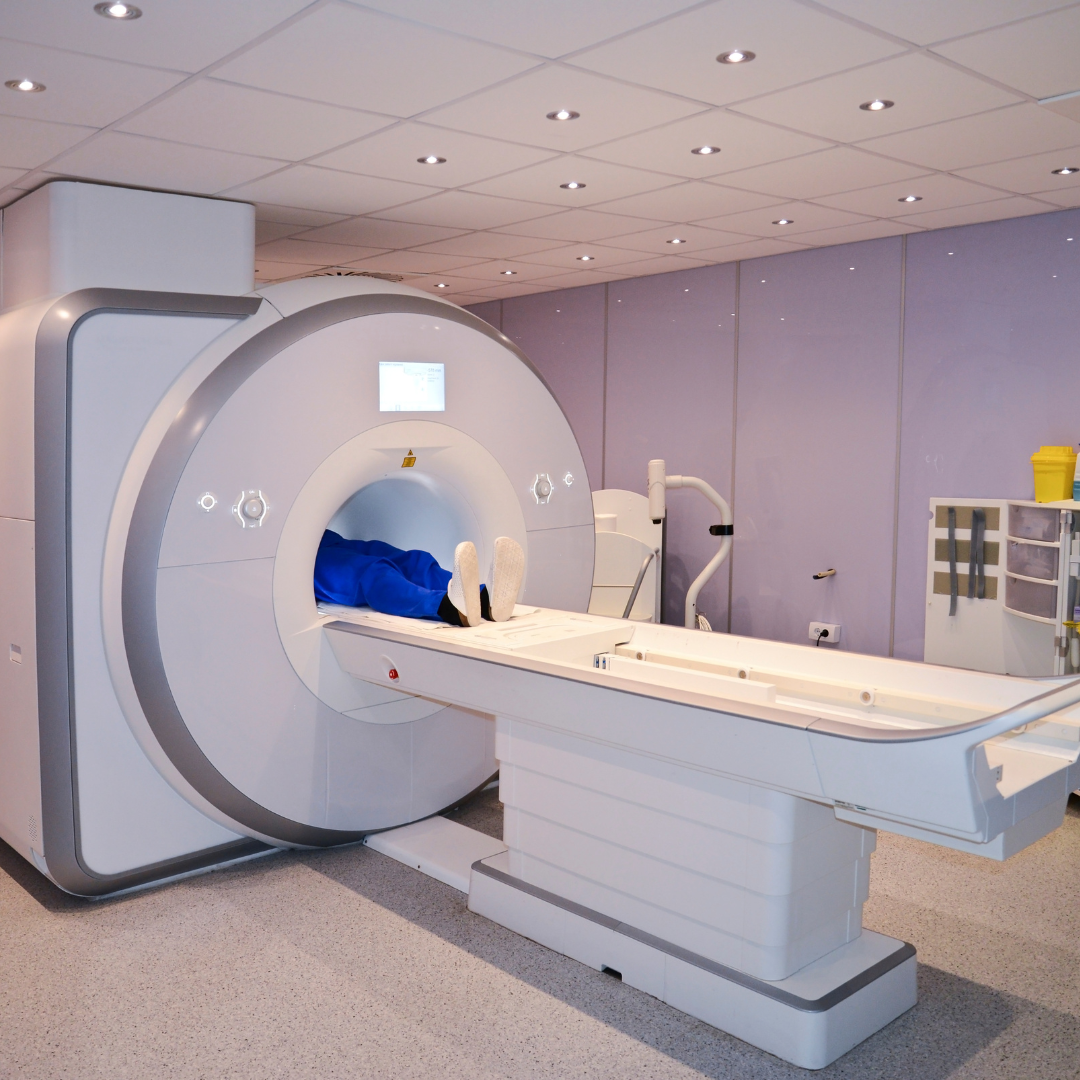
Hip and pelvis MRI without ionising X-ray radiation
MRI uses a strong magnetic field rather than X-rays, which makes it particularly suitable for the pelvis where radiation dose is a consideration. Because of the magnet, safety screening is essential before every scan.
Tell the imaging team about any implanted device, previous hip surgery or a hip replacement, metal fragments, or other relevant history. A hip replacement does not usually prevent a scan, but the radiographer will adjust the protocol and image quality near the metal is reduced. Read our full guide to MRI safety, implants and claustrophobia before you book.
Can you self-refer for a private hip or pelvis MRI?
New Horizons Imaging offers a self-referral pathway for selected non-emergency musculoskeletal MRI examinations, including hip and pelvis MRI, for adults aged 18 and over. Every request is reviewed by our clinical team before the scan is confirmed.
Pelvic MRI requested for gynaecological, prostate, bowel or other non-musculoskeletal reasons follows a different protocol and normally needs a clinician referral. If you already have a referral, upload it when you book. Patients under 18 always require one — find out how to get a referral here.
Hip and pelvis MRI at Headingley, Leeds
New Horizons Imaging provides private hip and pelvis MRI from AMT Headingley Rugby Stadium in Leeds on a Siemens wide-bore scanner, with free on-site parking and appointments often available within days.
Hip, pelvis and sacroiliac joint scans are each booked as a single body-area scan, and two areas can be combined in one visit. Current prices are on our MRI Services & Prices page, and you can read about what a private MRI costs and how long a scan takes before booking.
Patients travel to our Leeds MRI centre from across Yorkshire, including Bradford, Huddersfield, Sheffield, Halifax, Doncaster and York.
A routine private MRI appointment is not an alternative to emergency care. Inability to bear weight after a fall, a hot swollen joint with fever, sudden severe hip pain in a child or teenager, new numbness around the saddle area, or loss of bladder or bowel control should be assessed urgently rather than waiting for a routine scan.
Explore joint and sports MRI at New Horizons Imaging
Hip and groin injuries are among the most common in football, running and rugby. Our sports injury MRI guide explains how imaging fits into recovery planning, and our knee MRI and shoulder MRI guides cover the other joints we are most often asked about.
View our MRI Services & Pricing page for current scan options, or visit Areas We Serve if you are travelling into Leeds.
Unsure whether you need a hip, pelvis or spine scan? Contact the New Horizons Imaging team before booking.
Frequently asked questions about hip and pelvis MRI
What is the difference between a hip MRI and a pelvis MRI?
A hip MRI gives high-detail images of one hip joint — labrum, cartilage, bone and the tendons around it. A pelvis MRI takes a wider view of both hips, the pelvic bones, pubic symphysis, sacroiliac joints and muscle attachments. The right choice depends on where the symptoms are and what is suspected.
Can a hip MRI show a labral tear?
Yes. MRI is the main non-invasive test for labral tears and hip impingement. In some cases a surgeon may request an MR arthrogram, with contrast injected into the joint, for extra detail; this needs a clinician referral.
Will an MRI show hip arthritis?
Yes. MRI shows cartilage loss, bone changes and joint fluid associated with arthritis, and can detect early changes before they are visible on X-ray. X-ray remains useful for grading established arthritis and planning joint replacement.
How long does a hip or pelvis MRI take?
A hip MRI takes around 20 to 30 minutes of scanning and a pelvis MRI around 30 minutes. If both are requested together allow a little longer. See our guide on how long an MRI scan takes for more detail.
Do I need to prepare for a hip MRI?
No special preparation is needed for a musculoskeletal hip or pelvis MRI. Eat and drink normally, take usual medication, wear clothing without metal fastenings and complete the safety questionnaire when booking.
Can I have a hip MRI if I have a hip replacement?
Usually, yes. Modern hip replacements are MRI-safe, although the metal reduces image quality close to the implant. Tell us when booking so the radiographer can adjust the protocol, and read our MRI safety guide for details.
Can I self-refer for a private hip or pelvis MRI?
Adults aged 18 and over can self-refer for musculoskeletal hip and pelvis MRI at New Horizons Imaging, subject to clinical review. Pelvic scans for gynaecological, prostate or bowel reasons, and all scans for patients under 18, require a clinician referral.
Where does New Horizons Imaging perform hip and pelvis MRI scans?
New Horizons Imaging provides private MRI scanning from AMT Headingley Rugby Stadium, St Michael's Lane, Headingley, Leeds LS6 3BR, with free on-site parking.
Hip, groin or pelvic pain? Find out what's causing it.
If you know you need a hip or pelvis MRI, you can begin your booking online now. If you are unsure which scan is right, send us your referral or symptoms and our clinical team will confirm the examination before you pay.